The Dead Don’t Scare People; People Scare People

There’s no shortage of scareable moments in the morgue. I was unlucky enough to experience one during a recent shift.

Our machine was under repair and we had to use the portable for cases. Once positioned, I am too short to reach the controls on the x-ray tube. A smart x-ray tech would set the technique before positioning the tube, but let’s see how logical you are at 5 am.

As a side note, I should mention my PPE. PPE stands for personal protective equipment and is necessary when working with dead bodies. It includes a gown, two sets of gloves (I like to double up), a cap, a mask, and booties. The booties are important, I promise.

After standing on the tip of my toes and reaching, I decided to stop being lazy and get the foot stool. Conveniently, the stool is located in a separate room: the decomp room. The decomp room is a special room where the stinkiest of cases go. Fortunately, on that morning, no one was in there.

I opened the door and took in the creepiness of the room. It was dark and too quiet. I turned on one of the lights and began to walk across the room to where the foot stool was located. As I did, I heard, “psst”.

Frozen in my tracks, I listened carefully for another “psst”. I looked around the room and checked for life. There’s a bathroom in here, and many of the techs use it when they don’t want to go into the locker room. The bathroom was empty. So I looked behind me to see if someone had poked their head in after me. The door was still closed.

I decided that the smartest thing to do is not stand frozen in the middle of the room so I grabbed the stool and ran. You know that feeling you have when you run up the basement stairs not looking behind you out of fear that you might actually see something? Multiply that by five and that’s how I felt as I ran out of the room.

Safely back in the x-ray room, I laughed at myself for being as afraid as I was. I told my partner how creeped out I was and sufficiently creeped her out with my story. Terror, much like misery, loves company. But I’m a skeptic. Weird things happen around here all the time, and I’m usually the first one to find the rational explanation to ease nerves.

Regardless, this silly event had me rattled. No one was in the room. As far as I know, there isn’t a speaker in there so it’s not like someone could have said it over an intercom. Could it have been outside the room? I ran through all of the possible explanations and came up with nothing.

So with a deep sigh I hung my head in shame and looked down at my feet. Booties.

The PPE booties we wear are made of plastic. As you walk, they make a not-annoying-at-all noise. A noise that’s loud enough to hear down at the other end of the hallway.

Swish-swish, swish-swish, swish-swish, SWISH-SWISH.

The “psst” I heard was actually the “swish-swish” of my own feet.

Boom. Rationalized. Eerie feeling gone. I am an idiot.

My partner wasn’t as quick to accept my completely rational, totally logical founded-in-science explanation. So I walked around the x-ray room rubbing my feet together (swish-swish) while saying “psst psst,” in an effort to prove my theory.

She still doesn’t believe me.

I’m still an idiot.

Bullet Removal: That Was An Interesting Tactile Experience

When a homicide case comes in, particularly a gunshot, I cringe. Most likely not for the reason you’re thinking. My reaction has less to do with the loss of life and more to do with the fact that if the bullet didn’t exit the body, there’s a good chance this case will come back to me. Cold, I know, but the morgue hardens you.

An AP view of the skull with bullets and fragments. X-ray taken from a textbook.
A lateral view of the skull with bullets. X-ray taken from a textbook.

While it’s something I’m researching, I still don’t understand ballistics enough to go into the physics behind guns. What I can say is that whether the bullet exits a body has a lot to do with the bullet itself and where on the body the victim was shot.

I’m learning that bullet identification and recovery are important for several reasons. One, knowing the specific location of a bullet saves the pathologist a lot of time and minimizes needless searching during the autopsy.

Two, it’s important to carefully count the number of bullets present. They have to be correlated with the entrance and exit wounds, otherwise a discrepancy can lead to a search at the crime scene. It is possible that multiple bullets can enter through a single entrance wound, but typically only when automatic weapons are used.

Lastly, x-rays can reveal information about the angle and direction of fire. Small metallic fragments produced when a bullet strikes bone may lead directly to the bullet and clearly indicate the bullet’s path. This helps investigators recreate the positioning of the victim and their assailant.

**********************

And since it’s never safe to assume that there’s an exit wound, x-rays are mandatory.

The first thing I do is visually examine the body. I check the head, chest, arms, abdomen, and legs for any bullet holes. My partner does the same. Two sets of eyes are better than one. If we find any, then I make mental notes of areas that need to be more closely examined for bullets on the x-ray.

Then we scan the whole body using x-rays. Even if it seems that the victim had only been shot in the head, the bullet could have traveled into the neck or chest. Bullets frequently end up in a site that’s far away from their entrance points, especially if they’ve hit a bone, so it’s important to be thorough.

If the full body scan shows that there are bullets, I will often walk over to the body and try to feel for the them. Sometimes the bullet is superficial, meaning it’s just below the surface of the skin. They’re easily felt and sometimes they can even be seen as small bumps. If that’s the case, I put a piece of tape over the bullet and mark it with a “x”. The pathologists appreciate it because it makes for a very easy retrieval.

If the bullet isn’t easily palpable and is located within the skull, neck, chest, abdomen, or pelvis, then additional image needs to be taken. Specifically, a lateral view. Instead of x-rays going from the front side of the body to the back like in the AP view, the lateral lets you see left to right. It gives me and the pathologists an idea of whether the bullet is more anterior (toward the front of the body) or posterior (toward the back).

After I submit my x-rays and return the body to the cooler, I’m done with that case and it can now be autopsied. But despite my best efforts, bullets can be difficult to find. It could have shifted during the autopsy. I’ve seen bullets that appear to be in one area only to wind up in a lung that has already been removed from the body. I’ve also seen bullets, particularly those in the skull, get lodged within the sinuses. In the event of the latter, chances of the body being returned to x-ray are high.

I don’t like when bodies come back. By this point, the autopsy is underway and the chest cavity is open. Most if not all of the organs removed. The skull cap will also have been sawed off, skin peeled down over the facial bones, and brain removed. It’s a much different version of the body than I had previously worked with.

**********************

Recently I worked on a case where a man had been shot in the head. A large bullet fragment was lodged somewhere inside of the skull. The pathologist and tech tried to access it, but without x-ray, they were working blind. At this point, the body is returned to radiology and we use fluoroscopy to image the body.

Fluoroscopy is x-ray in real-time. When I take a regular x-ray, I’m given a static image in return. But with fluoroscopy, I can see a moving image. For example, if I were to stick my hand into the empty skull cavity, I could see my fingers wiggle in real-time.

When using fluoroscopy, forensic x-ray techs will often use a scalpel or tool with a long handle to help locate bullets. This keeps our hands out of the x-ray beam, minimizing our dose. I was using such a tool during this case, but our equipment wasn’t working very well. The image on the screen was very poor quality and I had a difficult time recognizing where I was within the skull.

The pathologist and her tech were anxiously awaiting my verdict, standing safely behind a lead-lined barrier. My partner, a new tech who had only recently joined the team at the morgue, was operating the C-Arm for me while I searched. I was starting to feel the pressure, so I decided to rely on my instincts. In order to do that, I had to ditch the tool and use my hand. This isn’t something you should do, as it increases your radiation exposure. However, I needed to feel the structures and I needed to find the bullet fast.

I saw my hand moving on the screen in front of me, inching closer to the dark spot: the bullet. I was hyper focused until I wasn’t. Something has pulled me out of the blissfully ignorant bubble I had put myself in. Suddenly I was very aware that my fingers were pushing along the ridges of facial bones and squeezing into holes that were inside of a human skull.

What the f*ck am I doing?

I had seen the other more seasoned x-ray techs do this dozens of time, but this was my first time as lead tech. There was no one else to ask. My new partner had just started and, to her, I was the seasoned tech.

“Is it stuck inside of the bone?”

I heard the doctor ask me a question, but clearly I had my hand inside of a man’s skull and couldn’t form a coherent response.

Sorry, I’m a bit busy not thinking about where my hand is right now. Okay, just focus on the screen. Feel the bones, identify the landmarks, find the bullet. Easy peasy.

After more palpating and a lot of internal shrieking, I felt it. I found the bullet. It was wedged within the sphenoid sinus. I grabbed my tool, used it to replace my finger, and called the pathologist over for removal.

I felt victorious, upset, super gross, and exhausted all at once. Thankfully it was near the end of my shift so a hot shower wasn’t too far off.

It’s a weird thing, being an x-ray tech in a morgue. On one hand, x-ray techs have clearly defined responsibilities. We’re taught to work within our scope of practice—provide exceptional patient care, minimize exposure to patients and personnel, evaluate images and ensure they’re of diagnostic quality. That’s the succinct version anyway. However, I’m learning that the lines which define our scope are blurred here. One day you’re taking a routine chest x-ray and the next you’re wrist deep inside of a dissected skull.

It’s Fine Until Someone Gets Slapped

When people find out where I work, their reaction is usually the same. First, it doesn’t completely register. “Oh, interesting.” And then the follow-up question: “What do you do there?” After I tell them that I x-ray human remains, it takes another second for that to sink in. “Oh. OHH!”

I’ll admit that this reaction is a little unusual to me, but that’s because I’ve spent so long researching this line of work that the connection just seems obvious. You work in the morgue, therefore you work with the dead. At least in some capacity.

What usually follows the “ah-ha” moment is one of the following statements:

  • “But you’re so small”
  • “But you’re so nice”
  • “But you’re so quiet.” (My personal favorite.)

I can see why people would think my size influences my ability to work with the dead, but my sound level? I don’t quite understand that one.

Even after all of that, a lot of people still don’t really understand what it is I actually do in the morgue. In this post, I’ll walk you through a typical day.

Elevator pitch: Got dead people? Then you need x-rays! Shave minutes off of autopsies with your very own set of radiographs! Locate bullets faster. Age fractures more accurately. Is that a nickel in the right lung?! No problem!

Heh.

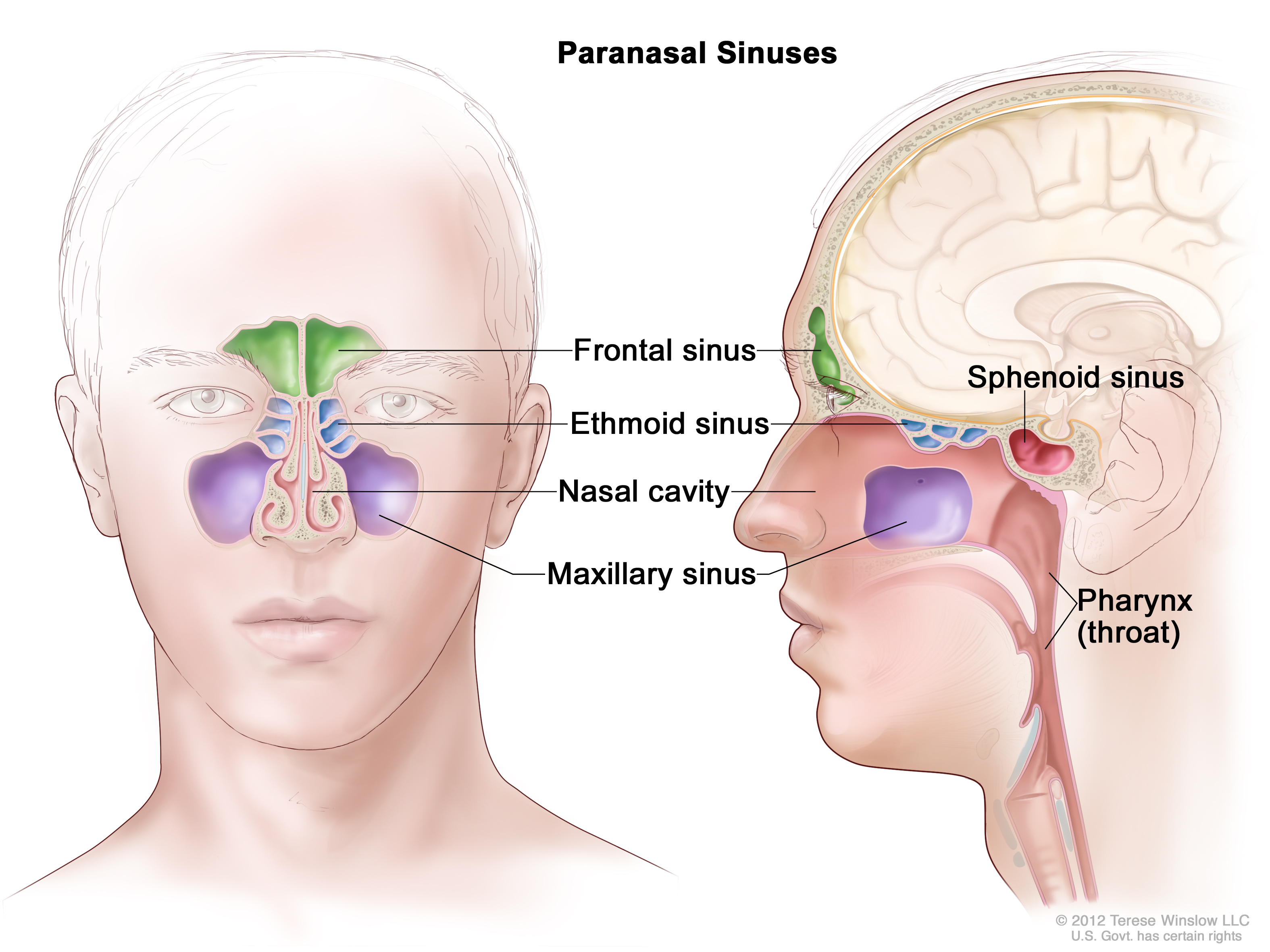
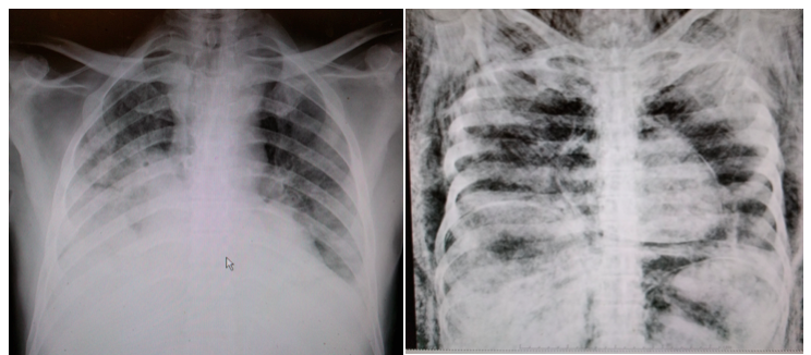
More accurately, I assist pathologists pre-autopsy by providing images of pathologies and traumas. X-rays offer a helpful roadmap in cases where foreign objects or bullets are involved. They also help determine the age of injuries: is this a new fracture or an old one? If new, did it contribute to the person’s death? Additionally, x-rays provide valuable legal documentation, which is important in homicide and abuse cases.

I’ve mentioned previously that we have certain protocols to follow when it comes to determining which bodies warrant x-rays. The first thing I do is read through all the investigation reports to learn the initial details surrounding an individual’s death. As I’ve said, every suspected homicide gets x-rays. This includes gunshot wounds and stabbings. Other cases needing x-rays include falls, motor vehicle accidents, drownings, fires, unknown bodies, and decomposing bodies.

We work in teams of two and each one has a specific role. One person is the “driver” and the other is the “runner”. As the driver, you operate the equipment and make sure everything is loading properly in the computer. You’re also responsible for annotating all of the images and keeping track of the number of x-rays taken and any bullets or foreign objects found.

As the runner, you’re responsible for tagging all of the bodies in the body cooler. This lets the pathologists and their techs know which individuals are going to x-ray. It also makes it easier for you to find the next case. Gunshot wounds take priority and get a red card, while everything else gets a yellow card.

I find this process unsettling at times. Usually it’s just you alone in the body cooler. At any given time, I’d estimate that there are close to 200 bodies in there. Many of them are being stored or awaiting transport to a funeral home. Only about 20-30 are actual cases for the day. You’re literally surrounded by death. It’s a heavy realization so early in the morning.

A co-worker likes to use this time to introduce herself to our cases. I’ve, on occasion, will say good morning to the cooler’s occupants. Sometimes talking while in there calms my nerves. Other times it makes me irrationally worried that I might hear a “good morning” in return.

The runner also transports the bodies to and from the x-ray room and positions the body for imaging. Obviously the runner is much more hands on with the bodies. This doesn’t usually bother me, but some cases are more difficult to stomach than others. More on that later.

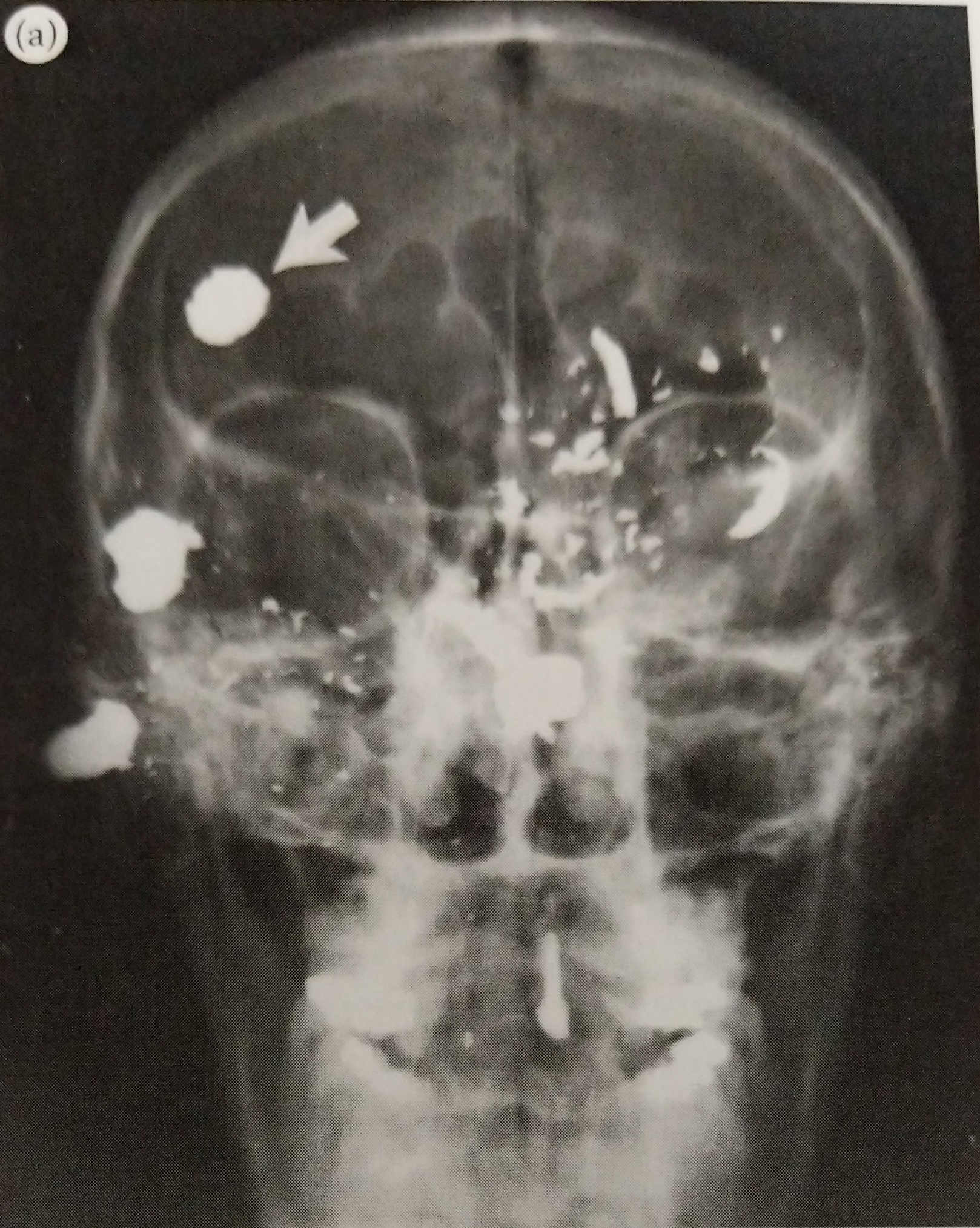
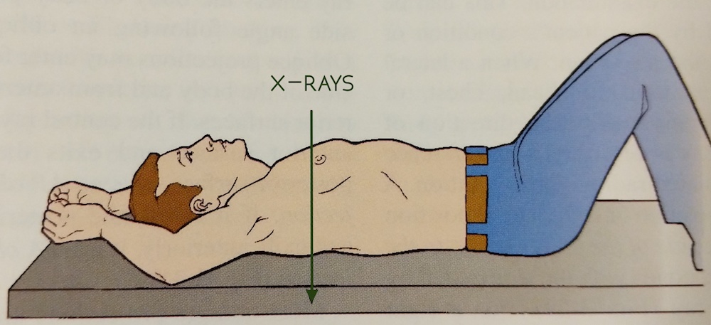
By default, every individual requiring x-ray will get at least one skull and one chest image. Most cases will get the standard protocol: skull, chest, abdomen, pelvis. Any area of trauma will also be imaged. In the event that the person is unknown, in addition to the standard protocol, we will take an x-ray of every long bone in the body: hands, forearms, arms, thighs, legs, and feet. All of these are performed with the body lying supine (on the back). These are called AP views.

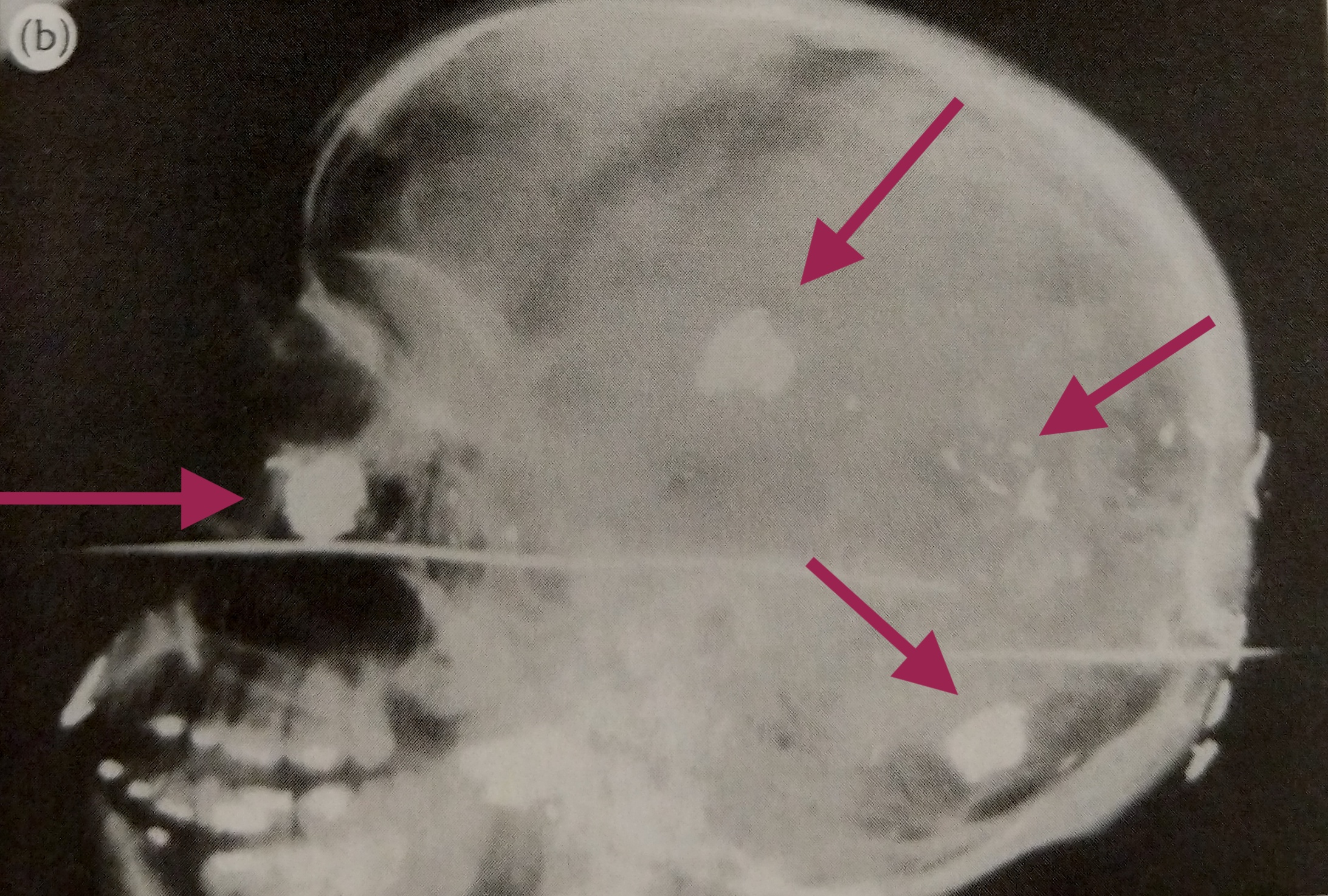
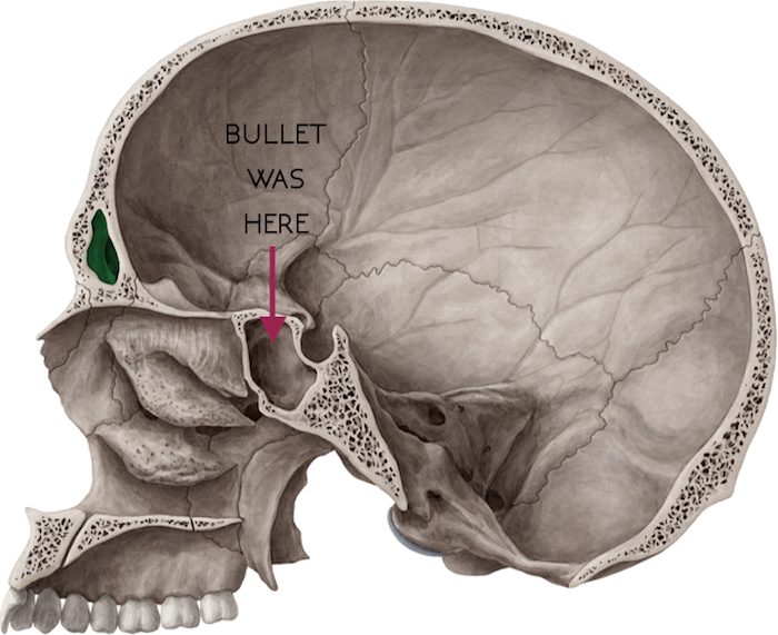
When there are bullets involved, we look for entrance and exit wounds. We cross our fingers for exit wounds because that means the bullet is no longer inside the body. This is good because we don’t have to do additional x-rays, and the pathologist doesn’t have to remove a bullet. But when there is no exit wound, we have to turn the body and get lateral views, meaning that the x-rays enter the right side of the body and exit the left side. This is different from the AP views, which enter the anterior (front side) and exit the posterior (back side).

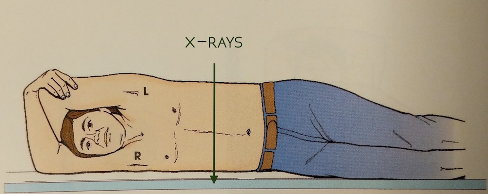
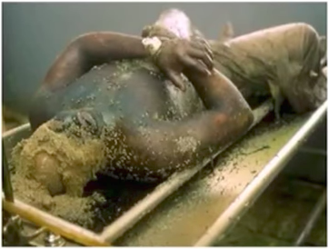
When you’re the runner, positioning for the lateral views falls on you. Most of the time it will require both technologists because many of the bodies are large. Let’s say someone was shot in the chest and they’re still holding a bullet. A lateral view will help a pathologist determine if the bullet is closer to the anterior or posterior surface. In order to get an unobstructed lateral view of the chest, the arms must be pulled above the head.

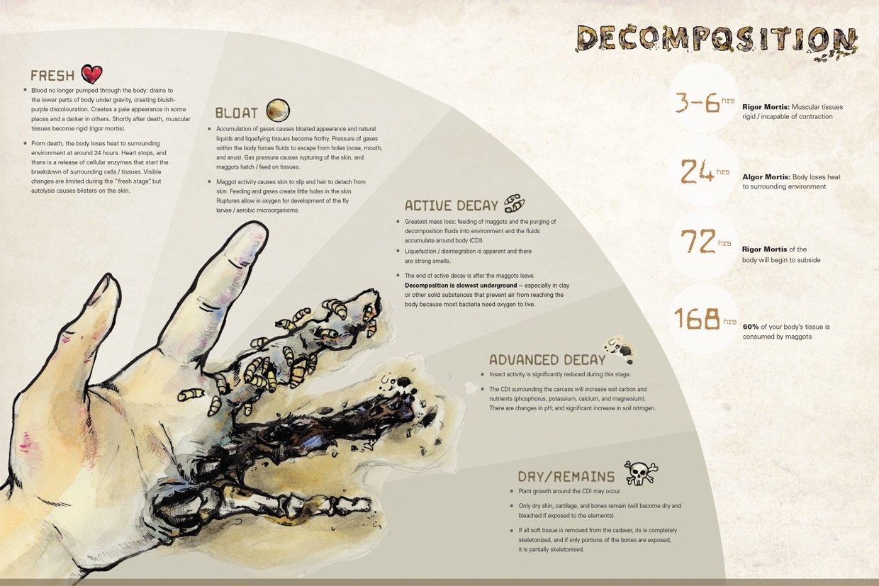
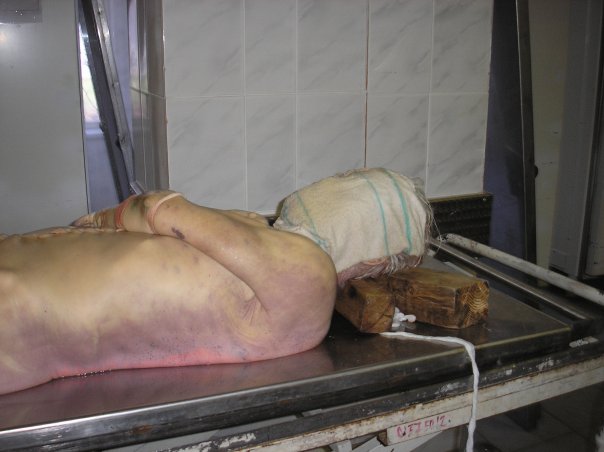
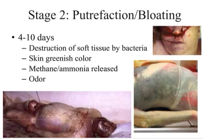
Did you read my post about decomposing bodies? If you did, then you know that most bodies on my x-ray table are in the rigor mortis stage of decomposition. This means that their muscles are severely contracted and moving extremities is very difficult. Nevertheless, it must be done. This is my least favorite part of the job. Not only do I feel like I’m always losing a very strange arm wrestling contest, but as the arm is pulled, there are cracks and pops. Occasionally you’ll dislocate a shoulder (theirs, not yours). And in cases where there is a lot of trauma to the arm, pulling it over the head can worsen the damage.

When the arms are successfully over head, they don’t always stay there. When you break rigor mortis, the muscles become more pliable. So when you put a body part somewhere, say an arm, it doesn’t always stay. Smart techs will use rope and sandbags to hold them in place. I? I am not a smart tech.

During a recent case, we had just taken a lateral chest x-ray. I was holding the body up on its side until the x-ray appeared on screen. You don’t want to move a body out of the lateral position until you’re sure you got the image. “All clear.” So I began to roll the body onto its back. As I did, the right arm began to leave its overhead position and swung down to slap me in the face.

F-word.

This is not unusual. In fact, it’s somewhat of an initiation for x-ray techs in the morgue. I hear the other is to have a body fall off the cart. I pray to the powers that be that it never ever happens to me.

Ever.

What made the slap worse was that it happened on the day I decided to take my mask off because every time I breathed it made my glasses foggy. Sigh.

Laterals are usually saved for last because of the likely chance that you’ll get a hand to the face. Or boob. Or ass. Death has no boundaries.

When we’re finishing up our x-rays, the pathologists are just starting their autopsies. We return to the radiology office and hang out until the autopsies are done, just in case a pathologist requests additional views or asks for help locating a bullet. When that happens, the pathologist will bring a body back to the x-ray room. I hate this because the body has already been dissected. Their chest and abdominal cavities are open, void of all their organs. Often times the skin around the skull has been pulled back and the brain is missing too.

It’s an odd feeling. Hours ago, there was a complete person on my table. Aside from whatever trauma they came in with, they were intact. They were whole. Now the shell is broken. The weight of the job is something I still struggle with. The separation of the person from the body doesn’t always happen instantaneously. Instinctually, I feel bad because the body is missing the parts that made it go: the heart and the brain. But I have a job to do so I must quickly flip the switch, disconnect, and see just a vessel. Not a person.

When all is done, we clean up the room. Any blood that reaches the floor stays there until the janitor comes through after our shift, but we wipe down the machines and our table so it’s ready for tomorrow’s guests. We return to our office, sometimes in silence, but usually in conversation, trying to shed the emotional weight. It’s weird. Some days are easier than others. In the beginning, it was difficult not to bring any my work home with me (not in the literal sense). I’d close my eyes while in bed and see the faces of the day’s cases. It was very unsettling.

Talking about it helps. Writing and working through my feelings is cathartic. I’m getting better at creating and enforcing emotional boundaries. Viewing it as a learning experience also helps. I don’t know how long I will be able to work at the morgue. It’ll be a constant challenge and will require a continuous balance of empathy and distancing. For now, it’s just too damn interesting to quit. So we’ll see what happens.

Why I’m Donating My Body to Science

“A donated organ can save a life, but a body provides the foundation to save many more.”

In 2009, my mom and I went to see Body Worlds, a traveling exhibit of dissected human bodies preserved through plastination. I had seen it before, but it’s always better to experience these things with another person. I wanted her to see why I was in such awe of the human body. I left the museum that day having made three decisions: my mom is a good sport, $30 for a organ donor t-shirt was totally worth it, and I am donating my body.

Up until that day, I hadn’t given much thought to what would happen to my body after I died. Do I want to be buried? Cremated? Shot into space? To be honest, none of that crossed my mind. I was 25 and invincible. But after walking through the Body Worlds exhibit for the second time, I knew that a coffin six feet under ground wasn’t for me. My atoms crave fame. (Not really. They crave caffeine.)

Body Worlds

Before leaving the exhibit, I used one of the computers there to sign up as a donator. Instead of donating my money, however, I opted to donate my body. It was almost too easy, and part of me believed that I had just sent one of those “wish you were here” museum postcards to a family member. But weeks later I received confirmation in the mail—I even got a fancy ID card to carry with me so people know what to do with my body when I die.

Side note: To be fair, it wasn’t just Body Worlds that had led me down this path. By this point in my life, I had attended several cadaver labs and read “Stiff” by Mary Roach. This book opened my eyes to not only the need for cadavers, but the very important purpose they serve in a number of research capacities. It’s a fascinating book, and she’s a brilliant author. I recommend reading it.

Nearly 10 years later, I haven’t changed my mind. My body will be donated when I die. What has changed is who I am donating it to. While the Body Worlds exhibit is enlightening, there isn’t much need for bodies to be plastinated and put on display. While it serves an educational purpose, it’s self-serving. What is needed, however, are bodies for medical students, anthropologists, ballistics experts, and first-responders.

Giant Heart

In 2016, National Geographic reported that the demand for cadavers is up, but the supply is down. The Anatomical Gift Association of Illinois reported that annual donations fell from 760 in 1984 to 520 in 2015. This was disheartening to read because body donation is such a wonderful gift. There are more than 20,000 enrolled first-year medical students, and for them, anatomy class is a rite of passage. With about six students assigned to one body, it really limits the amount of hands-on learning future doctors have.

According to NatGeo, in 2008, Colorado and Wyoming were 20 bodies short of the 158 cadavers requested by the states’ medical schools. And with physician assistant and nurse practitioner programs now utilizing cadavers, in addition to vocations outside of medical school, the supply is even more strained.

“There are more than 120 million registered organ donors in the United States, and an average of 79 people receive transplants each day, according to the U.S. Department of Health and Human Services. The federal government does not monitor whole body donations in the United States, but researchers estimate each year fewer than 20,000 Americans donate their bodies to medical research and training.”

This doesn’t sit right with me. I have gained so much knowledge from the generosity of body donors, and I’m not even in medical school. I don’t know what happens when we die, but I can’t imagine that I’d rest easily knowing that I opted to keep my body in a box instead of helping future medical professionals better understand anatomy, physiology, and pathology.

Now I’m not here to tell you what to do with your body. It’s yours. But I do hope that maybe I can answer some questions for you in the event that you’re considering body donation.

What is body donation?

Simply put, body donation is the donation of a whole body after death for education and research. Donated bodies are primarily used for medical education and research, but cadavers have helped industries outside of medicine, including NASA and car manufacturers.

What will happen to my body?

That all depends on where you donate your body. In medical settings, donated bodies are mostly used for gross anatomy and surgical anatomy. In 2015, Vice published an in-depth article about what happens to your body after it’s been donated. I recommend checking it out. But to be blunt, you will be dissected. Remember that fetal pig in high school biology? More than likely, though, you’ll be treated with much more respect than that pig.

Your body will be embalmed, which means your blood and other bodily fluids are replaced with chemical preservers. This makes it so your body will last instead of decompose. Side note: read my previous post all about decomposition.

In my personal experience with cadavers, I’ve worked with whole bodies and well as parts, such as arms and legs. I have seen skulls cut open to reveal the hollow space where the brain once sat. I have also seen heads split in half down the middle to reveal the inner workings of the nasal and pharyngeal areas. I have been the person doing the dissecting, and I’ve also been the person who observes the body post-dissection. It all depends on the class you’re taking and career path you’re heading down.

If you’re looking for a moving first-hand account of a cadaver lab, I recommend reading “Body of Work” by Christine Montross.

How can I donate my body?

There are several ways to do it. First and foremost, you can opt to be an organ donor. This is much more common than whole body donation. Working at the morgue, I receive a lot of Gift of Hope patients. These are bodies that have already had their corneas, organs, long bones, and even some skin removed.

My next recommendation is to check with your state to see if they have a formal organization. Illinois, where I am located, has the Anatomical Gift Association. It receives, prepares, preservers, and distributes donated human remains to medical education and research institutions across Illinois.

You might also consider research facilities like a “body farm”. The most notable one, at least in my opinion, is located at the University of Tennessee. It was founded in 1981 and has been used to study human decomposition. Similar projects have followed at Western Carolina University, Texas State University, Sam Houston State University, Southern Illinois University, and Colorado Mesa University. All six are, or at least at one point, accepted human donations. If you like podcasts, here’s a great one about body farms from the guys at Stuff You Should Know.

What happens to my body when they’re done?

That’s a great question. In my research, I’ve read that most places hold a type of memorial service or ceremony. Certain institutions will invite family members, while others prefer to limit it to the students and faculty that worked with the cadavers. It’s a great way to show respect and gratitude for the generous gift. Bodies are then cremated, and their remains are returned to their families.

So there you have it. It’s a very quick overview of body donation. If you are considering it, I encourage you to research further. Like me, you might change your mind about where you’d like your body to go or what you’d like it used for. It’s an important decision, and one that shouldn’t be made impulsively. That said, it’s truly a wonderful gift and one that I am extremely proud to give.

A Composition on Decomposition

I have worked at the morgue for a little over a month and I can say with certainty that the worst part of my job is getting hands-on with head traumas and decomposed bodies.

I will get to head traumas another time. Right now I’d like to focus on decomposition because while disgusting, it’s a truly fascinating process from a physiological standpoint. Before I dive into the specifics, here’s a bit of background in case you’re just tuning in.

At the morgue, it is my job to x-ray every decomposed body that comes in, regardless of how the person died. As I stated in a previous post, I’m responsible for x-raying all gunshot wounds, motor vehicle accidents, and child deaths, without question. I’ll read reports from falls and suicides. Depending on the nature of the event, we may or may not need to x-ray them. Decomposed bodies always get x-rayed without question.

decompLet’s talk a bit about decomposition—yay! I should note that I will be showing some images that might be unsettling or upsetting. View at your own risk.

When you die, your heart stops beating and your body’s cells stop receiving oxygen. Typically brain cells are the first to die, while bone and skin cells can survive for several days. Blood begins to drain from vessels and pools in the lower-lying portion of the body. So if a person dies lying on their left side, that side will appear darker while the right side appears more pale. This is called livor mortis, or lividity.

livor mortis
{source}

Side note: There are several “mortis” stages that a body goes through. The ones not covered in great detail here are pallor mortis, which is a pale or ashy appearance, and algor mortis, which is a change in body temperature. Algor literally translates to coldness.

Roughly three hours after death, rigor mortis sets in. Rigor mortis is simply a stiffening of muscles. In a living body, calcium ions flow into muscle cells and promote attachment between two muscle fibers, actin and myosin. Once attached, this causes the muscle to contract. A muscle remains in this state until ATP arrives to pump the calcium out of the cell. However, in a dead body, no oxygen means no ATP. Skeletal muscles will remain contracted until the muscles start to decompose.

Fun facts about rigor mortis:

  • It’s Latin for “stiffness of death”
  • Facial muscles stiffen before larger muscles
  • It can start anywhere from 10 minutes to several hours following death and can last up to three days (it’s greatly affected by temperature)

As the cells within a body begin to die, there is nothing maintaining their structural integrity, and so their membranes begin to break. As they do, they release enzymes that begin eating the cell from the inside out. This is usually referred to as autolysis, which means self-digestion. While this is happening, microorganisms and bacteria produce very unpleasant odors called putrefaction.

Additionally, the leaked enzymes produce many gases, including hydrogen sulfide, carbon dioxide, and methane. The pressure from these gases cause the body to bloat, which is the second phase of decomposition. The pressure can become so intense that some bodies can actually double in size. Bloat usually occurs around five days after death, according to an article I read.

Bloat
{YouTube}

Fun fact: Tongues and scrotums aren’t immune to bloating. Bloat is most common in the face, abdomen, and in males, the scrotum. One of the only good things about working with decomposed bodies is that we don’t have to open the body bag. However, this can make positioning a body for an x-ray more challenging since landmarks aren’t easily identifiable. One of the ways we “get our bearings” is to feel for facial landmarks. Usually we go for the nose and eyes, but depending on the stage of decay, sometimes the tongue is a dead (heh, pun) giveaway. The tongue expands so much that it protrudes from the mouth acting almost like a handle.

The tech I trained with opened every decomp bag so I could see the process in its various stages. Disgusted, I still looked because my sense of curiosity is a glutton for punishment. Before opening the bag, she would say, “Here, feel this.” Completely at a loss for what body part my hand might be touching, I’d ask her what it was. “That’s his scrotum.” I skeptically pulled my hand away because it felt more like a knee or elbow than soft tissue. But I was proven wrong when she opened the bag. The scrotum had such a buildup of pressure due to the gases within the body that it had no choice but to expand to double its original size.

Although a great way to startle a first-week forensic tech, this can actually be dangerous. The buildup of pressure combined with the loss of integrity of the decomposing skin could cause the body to rupture. So think twice before you go poking around decomposed bodies, okay?

Black
{YouTube}

By this point, the combination of feeding maggots, purged fluids, and cellular breakdown means the body has entered active decay. During this time, liquefaction of tissues becomes more visually apparent and extremely strong odors exist. If you think a fresh dead body smells bad, you’re gravely mistaken. I don’t know how to begin to describe the smell of a decomposing body.

One article attempted to do so, stating:

  • Cadaverine and putrescine smell like rotting flesh
  • Skatole has a strong feces odor
  • Indole has a musty, mothball-like smell
  • Hydrogen sulfide smells like rotten eggs
  • Methanethiol smells like rotting cabbage
  • Dimethyl disulfide and trisfulfide have a foul, garlic-like odor

Another article went the less scientific route, asking 20 people to describe the smell of death. I think this one is the most accurate:

“Rotten eggs, feces, and a used toilet left out for a month x 1,000.”

Working with bodies in active decay is very difficult. The vicious attack on one’s nose is frustrating enough, but the bodies are slippery too. The pressure from the gases forces fluids within the body to escape through open orifices, like the mouth, nose, anus, etc. And since we’re working through the body bag. The material of the bag paired with the slippery goop makes for a very frustrated x-ray tech.

Toward the end of active decay, when all of the body’s soft tissue has decomposed, hair, bones, and cartilage remain. Although decomposition begins almost immediately following death, it can take up to one year for a body to completely decompose to a skeleton. From there, it can take another eight to 12 years to decompose a skeleton. It depends on a variety of factors. Is the body buried in a dirt grave? Was it buried in a coffin? Was it embalmed? If not buried, what’s the temperature and environment like?

After active decay, the body enters something called butyric fermentation. While I’m not certain this is an official stage of decomposition, it’s something that occurs once the body has dried out and butyric acid accumulates. Butyric acid is present in human vomit, so you can imagine how awesome this stage smells. During this “stage,” the skin turns leathery and marks the transition from active decay to dry decay. In dry decay, roughly 50 days to one year after death, the body is almost completely dry and may actually mummify depending on environmental conditions.

At the morgue, I have worked with bodies in every stage of decay. None were pleasant, but I suppose the beginning stage is the easiest. When it comes to working with dead people, I prefer those that are still exhibiting rigor mortis. In a later post, you’ll see how I contradict that statement, but for now I stand by it. Without rigor mortis, bodies move too easily and seem too life-like. It interferes with the mental barrier I’ve constructed so I can do my job without puking and/or crying.

I debated whether or not I would show you this. I was given permission to use x-rays for education purposes at school, so I felt conflicted about posting anything here. I don’t think I’ll make a habit of this, but I really wanted you to see the differences between an x-ray of someone who had just died compared to that of someone who’s actively decaying.

Compare

Normal on the left and decay on the right.

Can you see the difference? Isn’t it interesting?! Because we work through the bags, I’m never quite sure what stage of decomposition the body is in. But the x-rays always show it. Now, an x-ray won’t tell you down to the day of decomposition the body is in, but it can give you a fairly good idea of whether it’s in the beginning stage or active stage. I think it’s fascinating. Hopefully, as I learn more about pathology and identifying different types on x-rays, I’ll be able to notice even the most subtle of decomposition markers. #XrayGoals